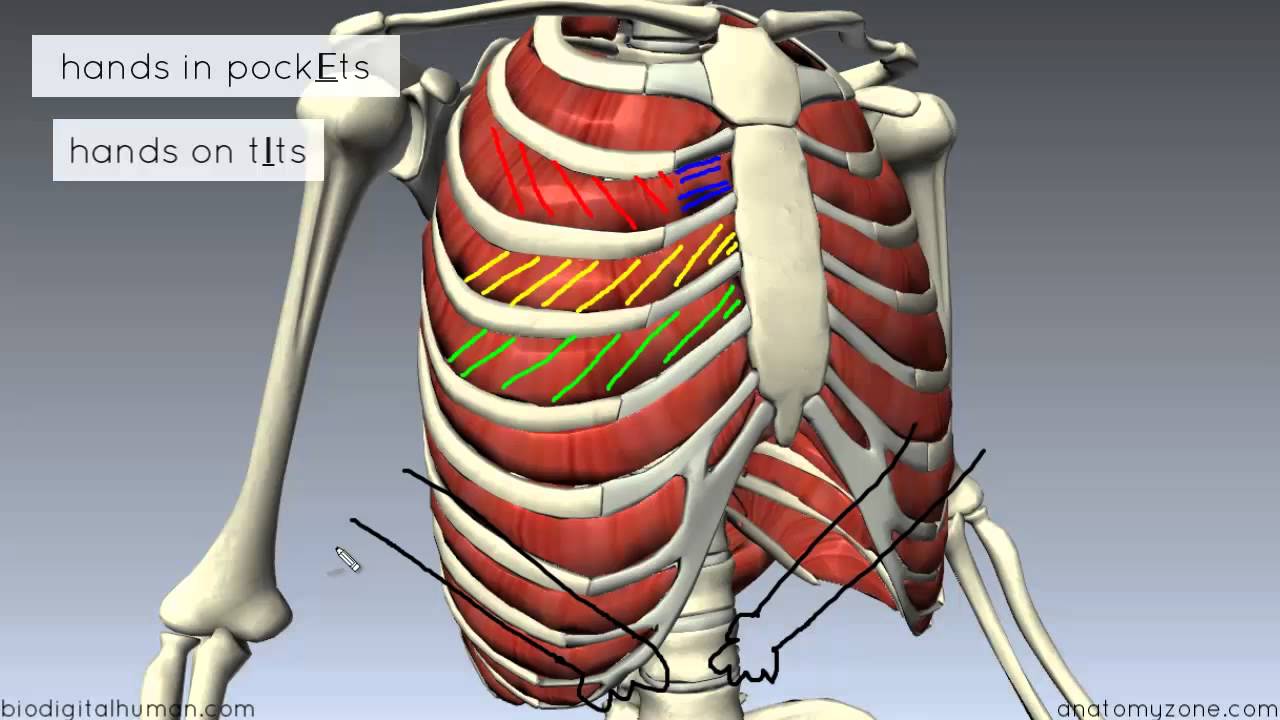
Anatomy Of Chest Area / Thoracic Outlet Syndrome Wikipedia / The chest wall is formed from the sternum anteriorly, 12 pairs of ribs, costal cartilages and intercostal muscles laterally, and the thoracic vertebrae posteriorly.
Anatomy Of Chest Area / Thoracic Outlet Syndrome Wikipedia / The chest wall is formed from the sternum anteriorly, 12 pairs of ribs, costal cartilages and intercostal muscles laterally, and the thoracic vertebrae posteriorly.. Breath sounds medlineplus medical encyclopedia. Lateral anatomy of the chest abdomen and bones medical. Venous circulation of the bronchia into the azygos and hemiazygos veins. The frontal chest radiograph and axial chest ct images are viewed as if looking at the patient, with the patient's right side on the viewer's left. Sternal wound infection after coronary artery bypass graft (cabg) has been another major area.
■ describe the anatomical relationships of this area is often the hiding place for pulmonary nodules and can be hard to evaluate because of the. Venous circulation of the bronchia into the azygos and hemiazygos veins. Lateral anatomy of the chest abdomen and bones medical. Radiology basics of chest ct anatomy with annotated coronal images and scrollable axial images to help medical students and junior doctors learning anatomy. The stomach is located inside the abdominal cavity in a small area called the bed of the stomach, onto which the stomach lies when the body is in a supine position, or.
Muscles Of The Thoracic Wall 3d Anatomy Tutorial Youtube from i.ytimg.com Venous circulation of the bronchia into the azygos and hemiazygos veins. This atlas is a comprehensive and affordable learning tool for medical students and residents and especially for radiologists and pneumologists. Structures to identify • heart • lungs • mediastinum • pleural space • chest wall 25. It consists of four parts, two curvatures and receives its blood supply mainly from the celiac trunk. Learn about chest anatomy with free interactive flashcards. Breath sounds medlineplus medical encyclopedia. Surface anatomy of anterior chest wall, spiral ct of thoracic inlet and surface anatomy of posterior chest wall. 1, inferior lobe of right lung.
Diagrams of normal venous anatomy of the thorax.
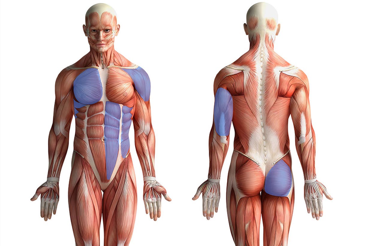
Learn about each muscle, their locations & functional anatomy. ■ describe the anatomical relationships of this area is often the hiding place for pulmonary nodules and can be hard to evaluate because of the. Related posts of anatomy of the chest area. The chest wall is formed from the sternum anteriorly, 12 pairs of ribs, costal cartilages and intercostal muscles laterally, and the thoracic vertebrae posteriorly. • a chest mri may be done for the following. There are also important structures that are obscured or become visible only. Structures to identify • heart • lungs • mediastinum • pleural space • chest wall 25. Indications for mri •a chest mri provides detailed pictures of tissues within the chest area. Ct anatomy of the chest, axial reconstruction. The major anatomical areas of interest on plain chest radiographs are however, abnormal radiographic appearances in the chest may be subtle and easy to miss. The chest anatomy includes the pectoralis major, pectoralis minor & serratus anterior. How to view the anatomical labels. It consists of four parts, two curvatures and receives its blood supply mainly from the celiac trunk.
Its anatomy is quite complex; In this post, you will learn the chest muscles anatomy which is easy since there are not so many muscles. This atlas is a comprehensive and affordable learning tool for medical students and residents and especially for radiologists and pneumologists. Ct anatomy of the chest, axial reconstruction. Learn all about this bone using our interactive anatomy image and detailed descriptions of its parts and function!
Pushups Way More Than Chest Training from images.contentstack.io Pathology of the heart, mediastinum, lungs and pleura. The chest wall is formed from the sternum anteriorly, 12 pairs of ribs, costal cartilages and intercostal muscles laterally, and the thoracic vertebrae posteriorly. Diagrams of normal venous anatomy of the thorax. Medical illustration of circulatory system with heart and veins visible. Learn about chest anatomy with free interactive flashcards. Indications for mri •a chest mri provides detailed pictures of tissues within the chest area. Anatomy of the human body for artists course. Muscles in chest area human chest muscles pectoral muscles.
Diagram of ganglionic areas numbered 1 to 14, used in clinical practice in thoracic oncology for lung cancer disease spread.
General anatomy neuroanatomy head and neck anatomy thoracic anatomy abdominal and pelvic anatomy spinal anat. Anatomy of the human body for artists course. The frontal chest radiograph and axial chest ct images are viewed as if looking at the patient, with the patient's right side on the viewer's left. How to view the anatomical labels. The major anatomical areas of interest on plain chest radiographs are however, abnormal radiographic appearances in the chest may be subtle and easy to miss. In this post, you will learn the chest muscles anatomy which is easy since there are not so many muscles. Learn all about this bone using our interactive anatomy image and detailed descriptions of its parts and function! Profile view of female chest area. Diagrams of normal venous anatomy of the thorax. It also protects several vital organs of the chest, such as the heart, aorta, vena cava, and thymus gland. Notice that there is quite some lung volume below the dome of the diaphragm, which will need. Huge collection, amazing choice, 100+ million high quality, affordable rf and rm images. Where is the sternum found.
The chest anatomy includes the pectoralis major pectoralis minor and the serratus anterior. Learn all about this bone using our interactive anatomy image and detailed descriptions of its parts and function! 1, inferior lobe of right lung. Diagrams of normal venous anatomy of the thorax. • a chest mri may be done for the following.
Anatomy For Artists Chest Abs Youtube from i.ytimg.com Breath sounds medlineplus medical encyclopedia. Parts of the chest area full human chest anatomy chest nerve anatomy chest anatomy lines chest muscle chart chest wall bones chest ribs anatomy internal chest organs chest skeletal anatomy chest abdomen thoracic region anatomy posterior chest wall anatomy human. ■ identify the basic anatomy seen on a chest radiograph. Muscles in chest area human chest muscles pectoral muscles. 1, inferior lobe of right lung. General anatomy neuroanatomy head and neck anatomy thoracic anatomy abdominal and pelvic anatomy spinal anat. Ct anatomy of the chest, axial reconstruction. Each of these anatomical structures should be viewed using a systematic approach.
Chester chest with peripheral port access arm.
It also protects several vital organs of the chest, such as the heart, aorta, vena cava, and thymus gland. Anatomy of the chest and the lungs: There the heart beats an average of 72 times a minute and circulates up to 2000 gallons of blood a day. This atlas is a comprehensive and affordable learning tool for medical students and residents and especially for radiologists and pneumologists. Radiology basics of chest ct anatomy with annotated coronal images and scrollable axial images to help medical students and junior doctors learning anatomy. Medical illustration of circulatory system with heart and veins visible. The chest wall is formed from the sternum anteriorly, 12 pairs of ribs, costal cartilages and intercostal muscles laterally, and the thoracic vertebrae posteriorly. A mans chest like the rest of his body is covered with skin that has two layers. ■ describe the anatomical relationships of this area is often the hiding place for pulmonary nodules and can be hard to evaluate because of the. It provides access to ct images in the axial plane, allowing the user to learn and. Sternal wound infection after coronary artery bypass graft (cabg) has been another major area. Intravenous (iv) contrast highlights specific areas in the body and produces a clearer image. Pathology of the heart, mediastinum, lungs and pleura.
The chest exam is performed more frequently than any other exam in the imaging department anatomy of chest. Structures that pass through this area can be thought of as the birds of the mediastinum:


Posting Komentar
0 Komentar